Physiological Effects of Nano L-carnitine on the Gonadal Pathway in Adult Male Rats Exposed to Lead Acetate
Abstract
This study was conducted to determine the role of L-Carnitine (LC) and Nano L-Carnitine protected male rats from testicular toxicity caused by lead acetate (PbAc) by investigating the effectiveness of biosynthesized L-Carnitine nanoparticles (LC-NPs) on the fertility status of male rats. LC-NPs were synthesized, which could be by encapsulating l-carnitine in Chitosan (CS)-tripolyphosphate (TPP)-based nanoparticles (NP);
Materials and methods: six groups of adult Wistar albino rats (10 per group) were divided as follows: The first group (control group): received orally Distilled water. the second group: received orally lead Acetate at a dose of 30 mg/kg of body weight daily for 30 days. third group: received (lead acetate 30mg/kg b.w.for 30 days + L-carnitine 100mg/ kg b.w. /day for two months). fouth group received lead acetate 30mg/kg b.w.for 30 days +Nano L-carnitine 100mg/ kg b.w./day for two months). fifth group: received L-Carnitine orally at a dose of 100mg/ kg b.w./day for two months. sixth group: received L-Carnitine-NPs orally at 100mg/ kg b.w./ day for two months.
Results: a significant increase (P<0.05) in the testosterone hormone levels in the lead acetate+ Nano L-Carnitine group compared with the lead acetate group. While showed decreased levels of FSH and LH in the lead acetate+ Nano L-Carnitine group compared with the lead acetate group.
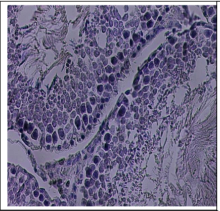
On the other hand, The results showed a significant increase (P<0.05) in the percentage of count and motility in group three(lead acetate+l-carnitine) and group four (lead acetate + nano l-carnitine) compared with group two (lead acetate). Show better improvements in the percentage in group four. Also showed a significant (P<0.05) decrease in the rate of dead sperm and abnormal morphology in group three and group four compared with group two (lead acetate). Histopathological changes were marked in testes; these changes included damage. Severe degeneration and sloughing of germinal epithelium with basement membrane disarrangement; no spermatid in seminiferous tubule lumens accompanied with atrophy and irregularity, vacuolization with few spermatogonia in seminiferous tubule lumens those changes reduced or disappeared in the treated groups.