Morphometric and Histological Study of the Papillary Muscles and Tendinous Chords of the Heart of Local Breed Bulls (Bos Taurus)
Abstract
This study was aimed to identify the anatomical and histological structures of papillary muscles of the right and left sides of bulls’ hearts, in relation to the chordae tendineae which are normally extended from papillary muscles. Six hearts of fresh adult bulls were studied grossly and histologically for this research.
Anatomical study was conducted by dissecting bulls’ hearts, and length, breadth of papillary muscles and number of chordae tendineae were described and measured. For histology study, special and routine stains were used to distinguish the general pattern contents of muscles, types of fibers and connective tissue. This study found that there were three papillary muscles in the right ventricle and two papillary muscles in the left ventricles, so the angular papillary muscle was absent in the left side. However, septal and parietal papillary muscles of left side were the largest papillary muscles. Also, the numbers of chordae tendineae were observed more numbers in the right papillary muscles.
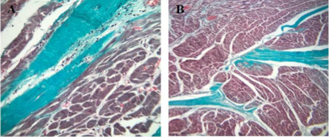
Histologically, myocytes were similar into cardiac cell muscles. Although, there are increasing the scattered more connective tissues and fibers. The chorea tendineae was no myocytes and completely transited into connective tissue.
Briefly, this study was indicated that angular papillary muscle are the largest muscle and absent in left side. However, more chordae tendineae were detected in right side, in addition, the myocytes were shorter than cardiac cells, but gradually, myocytes were disappeared at the chordae tendineae. This results in relation to pathological heart diseases could be explained more reasons for heart failure diseases.