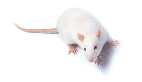
Hematological, Biochemical, and Histological Measures in Wistar Male Rats used to Assess Asafetida's Toxicity
Abstract
Traditional uses of asafetida in folk medicine include the alleviation of a wide range of symptoms. A proper assessment of its toxicity in the animal system is necessary to back up its usage in conventional medicine. Asafetida was tested for its toxicity in Wistar albino rats in order to draw conclusions about its safety for human consumption. Tools and Techniques: Animals were given oral doses of different mg/kg of body weight to test for acute toxicity. Asafetida was given to animals at several dosages of mg/kg body weight) during the course of a 6-week chronic trial. Asafetida's effects on renal,haematological, and histological indicators and hepatic measures were assessed at the study's conclusion. There was no fatality seen in the acute toxicity trial for asafetida up to 72 hours after dosing. Within 24 hours, no noticeable neurological or behavioural abnormalities were seen. In the long-term trial, asafetida consumption altered haematological markers including platelets, RBC, WBC, andHCT. The enzymes lactate dehydrogenase (LDH) and aspartate aminotransferase (AST)were both considerably elevated in the treated animals. At no point throughout the research did asafetida treatment cause a change in plasma urea or creatinine levels. Hepatotoxicity was found by histopathology analysis, although significant kidney pathological alterations were not. We conclude that asafetida is safe for short-term use, but that long-term use may have unfavourable effects on hepatocytes and haematological parameters.
References
Iranshahy M, Iranshahi M. Traditional uses, phytochemistry and pharmacology of asafoetida (Ferulaassa-foetida oleo-gum-resin) -a review. J Ethnopharmacol. 2011;134:1–10.
Zargari A. Medicinal Plants. Tehran: Tehran University Publications; 1996. pp. 529–602.
Emami A, Fasihi S, Mehregan I. Medicinal Plants. Vol. 1. Tehran: Andisheh Avar Publications; 2010. pp. 24–8. [Google Scholar]
Eigner D, Scholz D. The magic book of Gyani Dolma. Pharm Unserer Zeit. 1990;19:141–52. [PubMed] [Google Scholar]
Bagheri S, HejazianSh, Dashti-R M. The Relaxant Effect of Seed's Essential Oil and Oleo-gum-resin of Ferulaassa-foetida on Isolated Rat's Ileum. Ann Med Health Sci Res. 2014;4:238–41. [PMC free article] [PubMed] [Google Scholar]
Bagheri SM, Rezvani ME, Vahidi AR, Esmaili M. Anticonvulsant effect of Ferulaassa-foetida oleo gum resin on chemical and amygdala-kindled rats. N Am J Med Sci. 2014;6:408–12. [PMC free article] [PubMed] [Google Scholar]
Bagheri SM, Dashti-R MH, Morshedi A. Antinociceptive effect of Ferulaassa-foetida oleo-gum-resin in mice. Res Pharm Sci. 2014;9:207–12. [PMC free article] [PubMed] [Google Scholar]
Kelly KJ, Nue J, Camitta BM, Honig GR. Methemoglobinemia in an infant treated with the folk remedy glycerited asafoetida. Pediatrics. 1984;73:717–9. [PubMed] [Google Scholar]
Dr.Feroz Ahmad Shergojri, Dr. Savita. (2021). Reviewing Significance Of Biological Activities Present In Quinoxaline. Drugs and Cell Therapies in Hematology, 10(3), 338–342.
Dr.Feroz Ahmad Shergojri, Dr. Akanksha Anand Saxena. (2021). Studying Design Structure And Categories Of Antimicrobial Peptides (Amps). Drugs and Cell Therapies in Hematology, 10(3), 312–317.
Eigner D, Scholz D. Ferulaasa-foetida and Curcuma longa in traditional medical treatment and diet in Nepal. J Ethnopharmacol. 1999;67:1–6. [PubMed] [Google Scholar]
Kumar P, Singh DK. Molluscicidal activity of Ferula asafoetida, Syzygiumaromaticum and Carum carvi and their active components against the snail Lymnaea acuminata. Chemosphere. 2006;63:1568–74. [PubMed] [Google Scholar]
Ramadan NI, Abdel-Aaty HE, Abdel-Hameed DM, El Deeb HK, Samir NA, Mansy SS, et al. Effect of Ferulaassafoetida on experimental murine Schistosoma mansoni infection. J Egypt Soc Parasitol. 2004;34:1077–94. [PubMed] [Google Scholar]
Ramadan NI, Al Khadrawy FM. The in vitro effect of Assafoetida on Trichomonas vaginalis. J Egyp Soc Parasitol. 2003;33:615–30. [PubMed] [Google Scholar]
Sitara U, Niaz I, Naseem J, Sultana N. Antifungal effect of essential oils on in vitro growth of pathogenic fungi. Pak J Bot. 2008;40:409–14. [Google Scholar]
Garg DK, Banerjea AC, Verma J. The role of intestinal Clostridia and the effect of asafoetida (Hing) and alcohol in flatulence. Indian J Microbiol. 1980;20:194–7. [Google Scholar]
Bagheri SM, Sahebkar A, Gohari AR, Saeidnia S, Malmir M, Iranshahi M. Evaluation of cytotoxicity and anticonvulsant activity of some Iranian medicinal Ferula species. Pharm Biol. 2010;48:242–6. [PubMed] [Google Scholar]
Abraham SK, Kesavan PC. Genotoxicity of garlic, turmeric and asafetida in mice. Mutat Res. 1984;136:85–8. [PubMed] [Google Scholar]
Walia K. Effect of asafoetida (7-hydroxycoumarin) on mouse spermatocytes. Cytologia (Tokyo) 1973;38:719–24. [PubMed] [Google Scholar]
Devaki K, Beulah U, Akila G, Gopalakrishnan VK. Effect of Aqueous Extract of Passiflora edulis on Biochemical and Hematological Parameters of Wistar Albino Rats. Toxicol Int. 2012;19:63–7. [PMC free article] [PubMed] [Google Scholar]
Sanderson JH. Philips: An atlas of laboratory animal haematology. New York: Oxford University Press; 1981;1:150–55. [Google Scholar]
Clarke EG, Clarke ML. Landers veterinary toxicology in London. London: Bailliere Tindall; 1997;2:456–61. [Google Scholar]
Will's DE. Biochemical basis of medicine. Bristol: John Wright and Sons Ltd; 1985;1:45–66. [Google Scholar]
Appidi JR, Yakubu MT, Grierson DS, Afolayan AJ. Toxicological evaluation of aqueous extracts of Hermanniaincana Cav. Leaves in male wistar rats. Afr J Biotechnol. 2009;8:2016–20. [Google Scholar]
Akanji MA, Yakubu MT. Alpha tocopherol protects against metabisulphite- induced tissue damage in rats. J Biochem Mol Biol. 2000;15:179–83. [Google Scholar]
Al-Habori M, Al-Aghbari A, Al-Mamary M, Baker M. Toxicological evaluation of Catha edulis leaves: A long term feeding experiment in animals. J Ethnopharmacol. 2002;83:209–17. [PubMed] [Google Scholar]
Ramakrishnan S, Swami R. Text Book of Clinical Biochemistry and Immunology. Vol. 3. Chennai: T.R. Publications; 1995. pp. 245–75. [Google Scholar]
Odoula T, Adeniyl FA, Bello IS, Subair HG. Toxicity studies on an unripe Carica papaya aqueous extract: Biochemical and haematological effects in Wister albino rats. J Med Plants Res. 2007;1:1–4. [Google Scholar]


